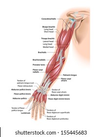
Diagram Of The Muscles In The Forearm - Forearm Wikipedia / The flexor pollicis longus is situated on the radial side of the forearm, lying in the same plane as the preceding.. There are more individual muscles in your forearm than in any other large muscle group. Editor · aug 11, 2017 ·. Longus, brevis, longus, brevis (longus is lateral to brevis). Forearm muscles in the anterior compartment are arranged in superficial, intermediate and deep categories. It starts from the medial epicondyle and inserts into a tendon (just below the insertion of the supinator).
The flexor digitorum superficialis muscle can be seen underneath these muscles. The muscles in the posterior compartment of the forearm are commonly known as the extensor muscles. Muscle anatomy diagram 12 photos of the muscle anatomy diagram canine muscle anatomy diagram, dog muscle anatomy diagram, lower leg muscle anatomy diagram, muscle anatomy of human back, tricep muscle. Try labeling diagrams and worksheets as additional learning aids. Diagram the movements of the humerus muscles that act on the forearm.
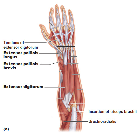
Bicep Muscle Diagram Images Stock Photos Vectors Shutterstock from image.shutterstock.com A very slight change in the length of the biceps causes a much larger movement of the forearm and hand, but the force applied by the biceps. The superficial extensors of the forearm are the brachioradialis, extensor carpi radialis longus, anconeus, extensor carpi radialis brevis, extensor carpi ulnaris, extensor digitorum and extensor digiti minimi. The forearm is the region of the upper limb between the elbow and the wrist. Try labeling diagrams and worksheets as additional learning aids. Learn vocabulary, terms and more with flashcards, games and other study tools. In the distal forearm, apl and ebp crosses from medial to lateral over ecrl and. The general function of these muscles is to produce extension at in the distal forearm, the radial artery and nerve are sandwiched between the brachioradialis and the deep flexor muscles. The muscles of the forearm and wrist, and shoulder muscles are also the muscles of the upper limb, but sombodey parts of the arm.
4, attachment… the muscles of the back forearm.
12 (4 superficial + 3 mobile wad + 5 deep). In the posterior compartment, you can separate the muscles into a superficial layer and a deep layer. 4, attachment… the muscles of the back forearm. So, the muscles of the anterior compartment are generally innervated by the median nerve, with a few muscles being innervated by the ulnar nerve. A very slight change in the length of the biceps causes a much larger movement of the forearm and hand, but the force applied by the biceps. Bend your palm toward your forearm. This layer contains only one muscle, the flexor digitorum. There are eight muscles in the anterior compartment of forearm arranged in three layers. Start studying muscles of the forearm. The forearm is divided into two compartments, which are separated by the radius and ulna and the interosseous membrane running between them. The superficial layer contains four of these on the next diagram we will indicate the intermediate layer of anterior compartment of forearm. Superficial muscles of the posterior forearm: The superficial extensors of the forearm are the brachioradialis, extensor carpi radialis longus, anconeus, extensor carpi radialis brevis, extensor carpi ulnaris, extensor digitorum and extensor digiti minimi.
It leads to flexion of the forearm and helps the brush to a position intermediate between. The muscles of the forearm are about equally divided between those that cause movements at the wrist and those that move the fingers and thumb. So, the muscles of the anterior compartment are generally innervated by the median nerve, with a few muscles being innervated by the ulnar nerve. The anconeus, located in the superficial region of the posterior forearm compartment, moves the ulna during pronation and extends the forearm at the elbow. The forearm is a mass of some 20 different muscles.
Muscles Of The Forearm from antranik.org All the muscles in the posterior compartment of the forearm are innervated by the radial nerve. Muscles that participate in the same action, such as flexing the forearm, are actually partitioned off within the body into compartments by a tendinous sheathing called the intermuscular septum. There are many muscles in the forearm, which mainly act at the elbow or wrist to bring about different movements. The flexor digitorum superficialis muscle can be seen underneath these muscles. The anterior forearm muscles are divided into 3 muscular layers ; Tutorials and quizzes on muscles that act on the forearm/ forearm muscles (flexors and extensors of the forearm), using interactive animations and diagrams. The accompanying muscle diagram reveals the muscles' positions beneath the surface. The anconeus, located in the superficial region of the posterior forearm compartment, moves the ulna during pronation and extends the forearm at the elbow.
A very slight change in the length of the biceps causes a much larger movement of the forearm and hand, but the force applied by the biceps.
The forearm is divided into two compartments, which are separated by the radius and ulna and the interosseous membrane running between them. The antibrachial or forearm muscles may be divided into a volar and a dorsal group. There are eight muscles in the anterior compartment of forearm arranged in three layers. Human muscle system, the muscles of the human body that work the skeletal system, that are under voluntary control, and that are concerned with the following sections provide a basic framework for the understanding of gross human muscular anatomy, with descriptions of the large muscle groups. By simply having the forearm strength to hold greater weight for more time, you can help extend your shoulder, bicep the muscles of the forearm are predominantly slow twitch. A deep layer , intermediate layer and superficial layer. The forearm is a mass of some 20 different muscles. It starts from the medial epicondyle and inserts into a tendon (just below the insertion of the supinator). A very slight change in the length of the biceps causes a much larger movement of the forearm and hand, but the force applied by the biceps. Editor · aug 11, 2017 ·. Bend your palm toward your forearm. The flexor pollicis longus is situated on the radial side of the forearm, lying in the same plane as the preceding. The muscles in the posterior compartment of the forearm are commonly known as the extensor muscles.
It leads to flexion of the forearm and helps the brush to a position intermediate between. The forearm is divided into two compartments, which are separated by the radius and ulna and the interosseous membrane running between them. Look at the picture of the muscle, find it on your body, and picture how it is contracting as it produces its associated movement or movements. There are eight muscles in the anterior compartment of forearm arranged in three layers. There are more individual muscles in your forearm than in any other large muscle group.
Muscles Of The Forearm And Wrist Diagram Quizlet from o.quizlet.com The general function of these muscles is to produce extension at in the distal forearm, the radial artery and nerve are sandwiched between the brachioradialis and the deep flexor muscles. This is the most medial of the superficial flexor muscles in the forearm. A helpful way to learn anatomy is to move and mimic the actions for the muscles you are learning that week. The flexor pollicis longus is situated on the radial side of the forearm, lying in the same plane as the preceding. Another handy relation to keep in the back of head is: Arm muscle diagram, forearm front arm muscle anatomy muscle diagram arm anatomy, anatomy of shoulder ligament ideas anatomy lesson full hd from the arm muscle diagram above, the muscles of the arm that can be seen easily on the surface include biceps, triceps, brachioradialis, extensor. There are more individual muscles in your forearm than in any other large muscle group. The forearm is divided into two compartments, which are separated by the radius and ulna and the interosseous membrane running between them.
The term forearm is used in anatomy to distinguish it from the arm.
Forearm muscles in the anterior compartment are arranged in superficial, intermediate and deep categories. This is the most medial of the superficial flexor muscles in the forearm. I've just switched over to a diagram to show you this muscle. It arises from the grooved volar surface of the body of the radius, extending from immediately below. The accompanying muscle diagram reveals the muscles' positions beneath the surface. The forearm is the region of the upper limb between the elbow and the wrist. Diagram of the muscles of the arm in action. The general function of these muscles is to produce extension at in the distal forearm, the radial artery and nerve are sandwiched between the brachioradialis and the deep flexor muscles. Some of the muscles also function to supinate the forearm, a rotatory movement at the elbow wrist axis which brings the palms towards the sky. The flexor pollicis longus is situated on the radial side of the forearm, lying in the same plane as the preceding. Diagram the movements of the humerus muscles that act on the forearm. The anterior forearm muscles are divided into 3 muscular layers ; Human muscle system, the muscles of the human body that work the skeletal system, that are under voluntary control, and that are concerned with the following sections provide a basic framework for the understanding of gross human muscular anatomy, with descriptions of the large muscle groups.
Diagram Of The Muscles In The Forearm - Forearm Wikipedia / The flexor pollicis longus is situated on the radial side of the forearm, lying in the same plane as the preceding.. There are any Diagram Of The Muscles In The Forearm - Forearm Wikipedia / The flexor pollicis longus is situated on the radial side of the forearm, lying in the same plane as the preceding. in here.